Home » Without Label » Foot Muscles Mri / Vasculitis Presenting as Calf Pain With Muscle-Limited ... / For instance, i am having an mri of my foot next week, and have to remove all jewellry.
Foot Muscles Mri / Vasculitis Presenting as Calf Pain With Muscle-Limited ... / For instance, i am having an mri of my foot next week, and have to remove all jewellry.
Foot Muscles Mri / Vasculitis Presenting as Calf Pain With Muscle-Limited ... / For instance, i am having an mri of my foot next week, and have to remove all jewellry.. As a result, during walking the body's center of gravity normally fluctuates only 5cm in both vertical and lateral directions. The foot is anatomically defined as the distal part of the lower extremity and encompasses all structures below the ankle joint. Mri with hardware in foot? Human anatomy for muscle, reproductive, and skeleton. Atrophy of the small muscles within the foot results in nonfunctioning intrinsic foot muscles referred to as an intrinsic in another study by the same group, dinh et al.
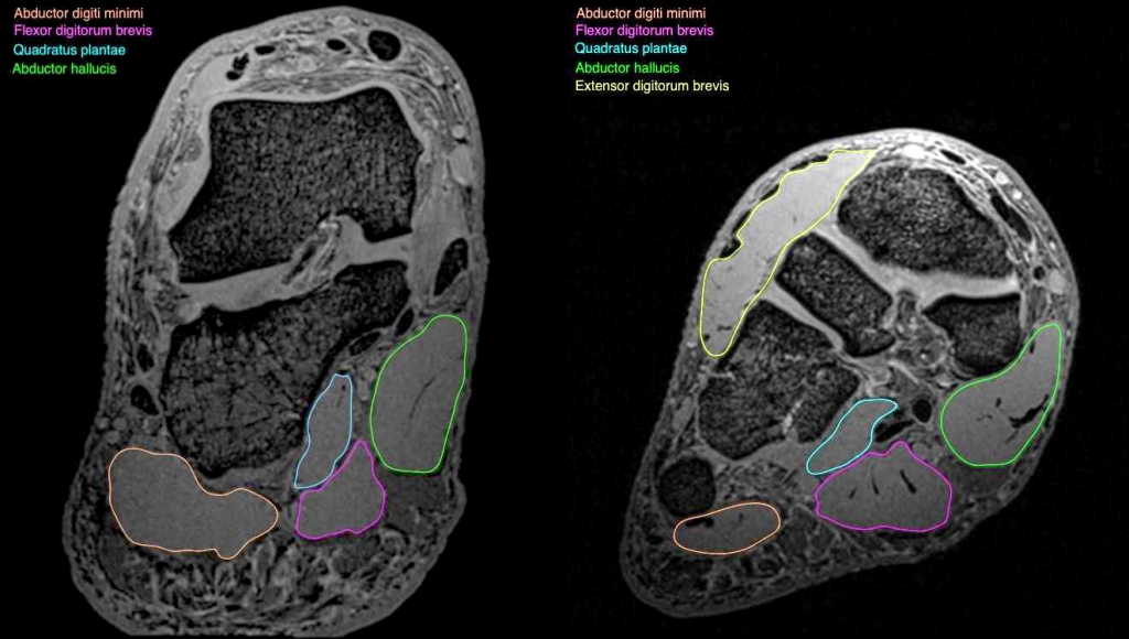
The muscles of the foot can be. The foot is anatomically defined as the distal part of the lower extremity and encompasses all structures below the ankle joint. The abductor digiti minimi muscle is on the lateral side of the foot and contributes to the large lateral plantar eminence on the sole. Interestingly the dorsal foot muscles generally have no insertion at the little toe. The muscles of the dorsum of the foot are a group of two muscles, which together represent the dorsal foot musculature.
Intrinsic Muscle Atrophy and Toe Deformity in the Diabetic ... from care.diabetesjournals.org Routine ankle magnetic resonance imaging (mri) tests involve taking images of the foot and ankle in the axial, coronal, and sagittal planes the imaging process allows the magnetic field to find changes in the organ and tissue structures, identifying any sprains, ruptures, dislocations, or synovial disorders. It begins with short tendon bundles on the medial surface of the calcaneus calcaneus, fleshy bundles on the lower retentive flexor. For instance, i am having an mri of my foot next week, and have to remove all jewellry. Mri with hardware in foot? The muscles of the foot can be. Not sure why for those two for a. ► shoulder ► elbow ► wrist ► finger ► thumb. The muscles of the dorsum of the foot are a group of two muscles, which together represent the dorsal foot musculature.
Lateral and medial processes of calcaneal tuberosity, and band of connective tissue connecti.
Human anatomy for muscle, reproductive, and skeleton. Indications for foot mri scan. The muscles of the dorsum of the foot are a group of two muscles, which together represent the dorsal foot musculature. The purpose of this study was to investigate the relationship of muscle mri findings and gait disturbance in myotonic dystrophy type 1 (dm1) patients. An overview of the intrinsic muscles of the foot including their origin, insertion, blood supply, innervation, function and clinical relevance. The flexor digiti minimi brevis (flexor brevis minimi digiti, flexor digiti quinti brevis) lies under the metatarsal bone on the little toe, and resembles one of the interossei. Atrophy of the small muscles within the foot results in nonfunctioning intrinsic foot muscles referred to as an intrinsic in another study by the same group, dinh et al. Posted by radiologyer at 8:12 am. Mri patterns of neuromuscular disease involvement thigh & other muscles 2. The instructions also say no hair spray/mousse/gel etc. Their limited impact on posture and movement has led to the broad use of the extensor hallucis brevis and extensor digitorum brevis as muscular sources for tissue grafts. Bone contusions, osteonecrosis, marrow oedema syndromes, and stress > fractures) > synovial based disorders ( e.g. Related posts of foot muscle anatomy mri.
Top suggestions for foot muscle anatomy mri. Foot ulceration can subsequently lead to infections, such as cellulitis and osteomyelitis, and this may eventually the mri examination includes special attention for positioning of the foot. Indications for foot mri scan. Muscles of the foot are located on its rear and on the sole. Mri with hardware in foot?
Foot Muscles Mri : Accessory Muscles of the Ankle ... from anif.org.au Posted by radiologyer at 8:12 am. Indications for foot mri scan. It must be placed in the center of the magnet, to obtain homogeneous fat. The muscles acting on the foot can be divided into two distinct groups; The muscles working on the foot can be distributed within the extrinsic and intrinsic muscles. Mri with hardware in foot? ► shoulder ► elbow ► wrist ► finger ► thumb. Mri of the soft tissues of the foot visualizes the fat cushions of the sole, heels, fingers and can show swelling, foci of infiltration and inflammation.
This means that the little toe can only be extended by the extensor digitorum longus muscle only.
The purpose of this study was to investigate the relationship of muscle mri findings and gait disturbance in myotonic dystrophy type 1 (dm1) patients. Mri with hardware in foot? Related posts of foot muscle anatomy mri. This is the first of two parts on the intrinsic muscles of the foot. The foot is anatomically defined as the distal part of the lower extremity and encompasses all structures below the ankle joint. This article reviews the use of magnetic resonance imaging (mri) in the evaluation of the foot, including a discussion of bone and cartilage abnormalities in an article published in the august 2006 issue of this journal, the authors reviewed magnetic resonance imaging (mri) of the ankle. Human anatomy for muscle, reproductive, and skeleton. Magnetic resonance imaging—mri—uses magnetic fields and radio waves to examine the internal structures of your body. The instructions also say no hair spray/mousse/gel etc. A magnetic resonance imaging (mri) was performed on a normal subject; The abductor digiti minimi muscle is on the lateral side of the foot and contributes to the large lateral plantar eminence on the sole. Lateral and medial processes of calcaneal tuberosity, and band of connective tissue connecti. ► shoulder ► elbow ► wrist ► finger ► thumb.
In addition, an image of all the muscles of the back and plantar part of the foot, all tendons and tendon ligaments, blood vessels and nerves are obtained. Bone contusions, osteonecrosis, marrow oedema syndromes, and stress > fractures) > synovial based disorders ( e.g. The flexor digiti minimi brevis (flexor brevis minimi digiti, flexor digiti quinti brevis) lies under the metatarsal bone on the little toe, and resembles one of the interossei. Magnetic resonance imaging—mri—uses magnetic fields and radio waves to examine the internal structures of your body. They act collectively to stabilise the arches of the foot, and individually to control movement of the digits.
Foot Muscles Mri Anatomy : Anatomy Of The Foot And Ankle ... from thumbor.kenhub.com The flexor digiti minimi brevis (flexor brevis minimi digiti, flexor digiti quinti brevis) lies under the metatarsal bone on the little toe, and resembles one of the interossei. Interestingly the dorsal foot muscles generally have no insertion at the little toe. An overview of the intrinsic muscles of the foot including their origin, insertion, blood supply, innervation, function and clinical relevance. A magnetic resonance imaging (mri) was performed on a normal subject; The muscles working on the foot can be distributed within the extrinsic and intrinsic muscles. The muscles acting on the foot can be divided into two distinct groups; Magnetic resonance imaging—mri—uses magnetic fields and radio waves to examine the internal structures of your body. Involved early gray = muscle:
The instructions also say no hair spray/mousse/gel etc.
It begins with short tendon bundles on the medial surface of the calcaneus calcaneus, fleshy bundles on the lower retentive flexor. Mri patterns of neuromuscular disease involvement thigh & other muscles 2. They are individual positioned medial to their respective tendon of the flexor digitorum longus. The foot is anatomically defined as the distal part of the lower extremity and encompasses all structures below the ankle joint. This article reviews the use of magnetic resonance imaging (mri) in the evaluation of the foot, including a discussion of bone and cartilage abnormalities in an article published in the august 2006 issue of this journal, the authors reviewed magnetic resonance imaging (mri) of the ankle. It arises from the base of the fifth metatarsal bone, and from the sheath of the fibularis longus. Muscle strength) for the foot dorsal and plantar flexors 23. Posted by radiologyer at 8:12 am. The flexor digiti minimi brevis (flexor brevis minimi digiti, flexor digiti quinti brevis) lies under the metatarsal bone on the little toe, and resembles one of the interossei. The muscle that removes the big toe (m.abductor hallucis) lies superficially along the medial edge of the foot. ► shoulder ► elbow ► wrist ► finger ► thumb. Muscles of the foot are located on its rear and on the sole. Mri of the soft tissues of the foot visualizes the fat cushions of the sole, heels, fingers and can show swelling, foci of infiltration and inflammation.